







5 to 7 percent job growth fuels demand for an ARRT MRI technologist skilled in advanced ECG gating and parametric mapping.
Table of contents
- Competition for jobs
- How to prepare for the future
- Skills that will matter more
- Advanced ECG gating and arrhythmia management
- Tissue characterization and parametric mapping
- Pharmacological stress testing and patient safety
- AI-accelerated imaging and compressed sensing
- Summary: is a Cardiac MRI Technologist career future-proof?
Cardiovascular magnetic resonance is moving from specialized academic research into mainstream hospital workflows. Cardiologists increasingly rely on MRI scans to assess heart failure, viability, and congenital anomalies, which creates steady demand for technologists who can manage complex cardiac protocols.
Competition for jobs
Group | Competition | Comment |
|---|---|---|
New graduates | Moderate | Entry-level technologists without dedicated cardiac training face competition for hospital-based cardiac scanner rotations. |
Experienced (ARRT certified, 3+ years cardiac) | Very low | High clinical volume and a scarcity of qualified operators keep certified ARRT MRI technologist specialists in short supply. |
How to prepare for the future
Technological shifts and faster scanning sequences require technologists to expand beyond routine anatomical imaging.
Earn specialized credentials: Complete Level 1 or Level 2 technologist verification through the Society for Cardiovascular Magnetic Resonance to validate your clinical competency.
Master hemodynamic monitoring: Learn to recognize arrhythmias, interpret real-time ECG waveforms, and adjust trigger delays during active scanning.
Adopt accelerated reconstruction tools: Practice working with AI-assisted k-space sampling and compressed sensing to manage patient breath-holds effectively.
Skills that will matter more
According to employment data from the U.S. Bureau of Labor Statistics, overall employment for radiologic and MRI technologists is projected to grow 5 to 7 percent through 2034 and 2035. The baseline requirements for scanning are expanding toward advanced physiological synchronization and rapid protocol adaptation.
Type of skill | Examples | Why it matters |
|---|---|---|
Hard (technical) | ECG gating, parametric mapping (T1/T2), 4D flow acquisition, stress agent safety | Clinicians require quantitative tissue characterization rather than purely visual assessments. |
Soft (clinical communication) | Breath-hold coaching, claustrophobia de-escalation, rapid team coordination | Uncooperative breathing or patient distress degrades image quality and lengthens table time. |
Advanced ECG gating and arrhythmia management
Cardiac imaging requires synchronizing pulse sequences with cardiac cycles to avoid severe motion artifacts. Technologists must decide when to use prospective triggering versus retrospective gating based on the patient's heart rate stability.
Prospective triggering initiates image acquisition at a predetermined point after the R-wave, making it suitable for flow quantification and targeted phases. The primary limitation is missing late diastole if the cardiac cycle varies.
Retrospective gating records image data continuously alongside the ECG signal, allowing reconstruction of the entire cardiac cycle for functional cine loops. Irregular rhythms like atrial fibrillation require real-time vector gating adjustments or arrhythmia rejection windows.
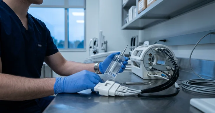
Electrode placement and lead selection determine signal quality inside high-gradient magnetic fields. Technologists must reposition leads, use vector loops, or switch to peripheral pulse gating when magneto-hydrodynamic voltage distortions obscure the true R-peak.
Tissue characterization and parametric mapping
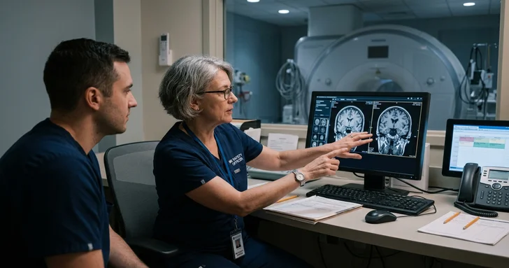
Modern cardiac exams have moved past qualitative scar identification to numerical tissue evaluation. Technologists must execute precise sequence timing and calibrate reference maps to measure myocardial properties accurately.
T1 mapping calculates absolute longitudinal relaxation times across myocardium to detect diffuse fibrosis and amyloid deposits. Technologists must run native maps before contrast administration and repeat sequences with matched parameters post-contrast to calculate extracellular volume fractions.
T2 and T2* mapping measure edema in acute myocarditis and quantify myocardial iron overload. Techs use standardized regions of interest and multi-echo gradient sequences to ensure reproducible decay curves.
Late gadolinium enhancement (LGE) reveals focal necrosis and infarction. Setting the inversion time (TI) accurately using TI scout or phase-sensitive inversion recovery (PSIR) sequences determines whether non-viable tissue is clearly delineated from normal myocardium.
Pharmacological stress testing and patient safety
Cardiac MRI stress protocols evaluate myocardial ischemia using vasodilators or inotropic medications. Technologists coordinate closely with attending cardiologists and nursing staff to ensure patient safety while capturing transient first-pass perfusion.
Vasodilator administration typically utilizes regadenoson or adenosine to induce hyperemia. Technologists time the dynamic contrast bolus during peak stress, requiring exact synchronization between power injectors and fast gradient-echo imaging sequences.
Implantable device safety involves screening and configuring MRI-conditional pacemakers and defibrillators. Technologists verify programming parameters, monitor real-time telemetry, and maintain specific absorption rate (SAR) limits within safety envelopes during scans.
AI-accelerated imaging and compressed sensing
Machine learning reconstruction algorithms are transforming scan times and breath-hold demands in cardiac suites. These tools reduce acquisition times from long multi-breath-hold exams to single-heartbeat or free-breathing acquisitions.
Deep learning reconstruction removes noise and sharpens borders on undersampled k-space data. ARRT MRI technologists must evaluate AI-reconstructed cine loops for unnatural smoothing or hallucinated anatomical edges before sending datasets to PACS.
Compressed sensing cine allows real-time cardiac function evaluation for dyspneic or pediatric patients who cannot hold their breath. Technologists adjust acceleration factors to balance temporal resolution against image sharpness.
Summary: is a Cardiac MRI Technologist career future-proof?
Yes. The rising prevalence of cardiovascular disease, combined with MRI's role as the reference standard for structural and functional heart assessment, ensures robust job security. Technologists who combine advanced gating proficiency with quantitative imaging expertise remain among the most protected and well-compensated professionals in medical imaging.
Tips if you want to enter the field:
Secure core credentials: Obtain primary or post-primary certification as an ARRT MRI technologist.
Gain supervised cardiac exposure: Pursue clinical rotations or training under an SCMR-recognized Level 2 or Level 3 center.
Master post-processing software: Familiarize yourself with cardiac analysis platforms like Circle cvi42, Argus, or Medis to assist with ventricular volume indexing and flow calculations.
Frequently asked questions
Yes. The rising prevalence of cardiovascular disease and MRI's role as the reference standard for heart assessment provide strong job security. Technologists with expertise in advanced gating and quantitative imaging remain in high demand.
Prospective triggering begins data acquisition at a set point after the R-wave, making it suitable for flow quantification. Retrospective gating continuously records data alongside the ECG signal, allowing reconstruction of the entire cardiac cycle for functional cine loops and better handling of irregular rhythms.
Key technical skills include advanced ECG gating and arrhythmia management, parametric tissue mapping (T1, T2, and T2*), late gadolinium enhancement (LGE) optimization, pharmacological stress protocol execution, and AI-accelerated reconstruction workflows.
Technologists can enter the field by securing ARRT MRI certification, pursuing specialized Level 1 or Level 2 technologist verification through the Society for Cardiovascular Magnetic Resonance (SCMR), gaining supervised clinical experience, and learning cardiac post-processing software.

Content manager
Erica Niklasson