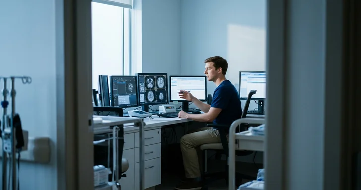








$95,480 per year is the median wage for MRI technologists. Research MRI technologists run 7T systems and execute custom neuroimaging protocols.
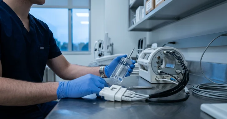
Table of contents
- Competition for jobs
- How to prepare for the future
- Skills that will matter more
- Ultra-high-field system operation (7T and above)
- Advanced neuroimaging and functional pulse sequences
- Deep learning reconstruction and protocol optimization
- Research safety compliance and data governance
- Summary: is a Research MRI Technologist career future-proof?
Research MRI technologists operate at the intersection of medical imaging and experimental science. According to the Bureau of Labor Statistics, employment for MRI technologists is projected to grow 5 percent through 2035, with a median annual wage of $95,480 recorded in May 2025.
Competition for jobs
Group | Competition | Comment |
|---|---|---|
New graduates | Moderate to high | Most academic medical centers require clinical experience and ARRT credentials before trusting technicians with experimental scanners. |
Experienced (5+ years) | Very low | An experienced ARRT MRI technologist with high-field physics or neuroimaging protocol background is rare and heavily recruited. |
How to prepare for the future
Working in research imaging requires technical adaptability beyond routine diagnostic scanning.
Master research protocols: Gain hands-on exposure to non-standard pulse sequences like functional MRI (fMRI), MR spectroscopy, and diffusion tensor imaging (DTI).
Earn advanced credentials: Maintain active ARRT MRI certification and pursue MR safety officer (MRSO) credentials through the American Board of Magnetic Resonance Safety.
Understand data pipelines: Learn how scanner data converts to research formats such as DICOM, NIfTI, and BIDS for analysis by principal investigators.
Skills that will matter more
Routine clinical workflows are increasingly automated by software, which pushes research technologist responsibilities toward protocol execution and hardware oversight.
Type of skill | Examples | Why it matters |
|---|---|---|
Hard (technical) | 7T ultra-high-field scanning, pulse sequence adjustments, physiological monitoring | Research centers use stronger magnetic fields and experimental sequences that require manual parameter adjustments and strict safety control. |
Soft (social) | Investigator communication, participant reassurance | Technologists must bridge the gap between academic research questions and participant tolerance during long scan sessions. |
Ultra-high-field system operation (7T and above)
Ultra-high-field systems provide high signal-to-noise ratios for brain and musculoskeletal research, but they introduce physical constraints absent in standard 1.5T and 3T systems. Operating at 7T requires managing dielectric shading, radiofrequency power deposition, and localized specific absorption rates (SAR).
B1 shimming and parallel transmit (pTx): Technologists configure multi-channel transmit coils and calibrate RF pulses to ensure uniform excitation across the field of view. Standard tools include vendor-specific shimming packages from Siemens Healthineers and GE HealthCare.
Acoustic and sensory management: Higher field strengths cause increased acoustic noise and subject dizziness from static field gradients. Technologists use specialized active noise-canceling headsets, visual display mirrors, and slow table-docking protocols to keep human subjects stable.
Advanced neuroimaging and functional pulse sequences
Clinical scans focus on standard anatomical contrasts, while research projects collect dynamic physiological and functional data. Technologists configure custom sequences designed to measure blood flow, metabolite concentrations, or tissue microstructure.
Functional MRI (fMRI): Technologists synchronize gradient-echo echo-planar imaging (EPI) sequences with external sensory cues, visual stimuli, and response pads. Software tools like E-Prime and PsychoPy trigger scanner acquisitions down to millisecond intervals.
Diffusion and spectroscopic imaging: Setting up high angular resolution diffusion imaging (HARDI) or multi-voxel MR spectroscopy requires precision in placement and fat-suppression tuning. Technologists evaluate real-time raw data to spot eddy current distortions or motion artifacts before concluding a session.
Deep learning reconstruction and protocol optimization
Deep learning reconstruction algorithms reduce scan times, but they require validation against raw k-space data in research settings. Technologists verify that machine learning reconstructions preserve subtle structural details without generating synthetic artifacts.
AI reconstruction engines: Platforms like Siemens Deep Resolve and GE AIR Recon DL process image data directly at the scanner console. Technologists monitor signal fidelity when testing these algorithms against proprietary research patches.
Artifact analysis: Technologists distinguish between true tissue signals and algorithmic reconstruction errors during novel sequence testing. This feedback helps imaging physicists refine mathematical reconstruction pipelines.
Research safety compliance and data governance
Research scanning often involves non-standard implants, experimental coils, and external physiological monitoring gear. The ARRT MRI technologist enforces strict Zone IV safety limits while keeping complete audit logs for institutional review boards (IRBs).
Non-standard hardware screening: Technologists evaluate custom RF surface coils, EEG caps, and eye-tracking cameras for magnetic displacement and heating risks. Testing follows ASTM International safety standards for medical devices in magnetic resonance environments.
Data de-identification: Technologists strip protected health information from image headers before transferring datasets to central repositories. They format files using standardized BIDS conventions for public data sharing and multi-center clinical trials.
Summary: is a Research MRI Technologist career future-proof?
Yes. As routine clinical imaging becomes automated, research facilities rely heavily on skilled technologists who understand pulse physics, safety at high field strengths, and study execution.
Tips if you want to go for it:
Target academic centers: Seek employment at university medical centers and teaching hospitals that maintain dedicated research scanners.
Build physics literacy: Study k-space trajectory, RF excitation, and gradient performance beyond standard board exams.
Master research software: Learn basic command-line utilities and image conversion tools to assist research teams with data workflows.
Frequently asked questions
Yes. While routine clinical workflows are increasingly automated, research facilities rely heavily on skilled technologists to manage complex pulse physics, execute non-standard protocols, oversee experimental hardware, and maintain safety at ultra-high magnetic field strengths.
Essential qualifications include active ARRT MRI certification, hands-on mastery of advanced neuroimaging protocols (such as fMRI, DTI, and MR spectroscopy), MR Safety Officer (MRSO) credentials, and knowledge of scientific data conversion pipelines like DICOM, NIfTI, and BIDS.
Employment for MRI technologists is projected to grow 5 percent through 2035, with a median annual wage of $95,480. While competition is moderate to high for new graduates, experienced technologists with neuroimaging and high-field physics experience face very low competition and are heavily recruited.
Operating at 7T and above requires managing dielectric shading, radiofrequency power deposition, and localized specific absorption rates (SAR). Technologists must also calibrate multi-channel transmit coils via B1 shimming and manage subject side effects such as static-field dizziness and elevated acoustic noise.

Content manager
Erica Niklasson